Xenon, Xe »
PDB 1w53-3pk4 »
2w6w »
Xenon in PDB 2w6w: Crystal Structure of Recombinant Sperm Whale Myoglobin Under 1ATM of Xenon
Protein crystallography data
The structure of Crystal Structure of Recombinant Sperm Whale Myoglobin Under 1ATM of Xenon, PDB code: 2w6w
was solved by
A.E.Miele,
F.Draghi,
F.Renzi,
G.Sciara,
K.A.Johnson,
B.Vallone,
M.Brunori,
C.Savino,
with X-Ray Crystallography technique. A brief refinement statistics is given in the table below:
| Resolution Low / High (Å) | 20.00 / 1.99 |
| Space group | P 6 |
| Cell size a, b, c (Å), α, β, γ (°) | 90.626, 90.626, 45.505, 90.00, 90.00, 120.00 |
| R / Rfree (%) | 18.8 / 20.5 |
Other elements in 2w6w:
The structure of Crystal Structure of Recombinant Sperm Whale Myoglobin Under 1ATM of Xenon also contains other interesting chemical elements:
| Iron | (Fe) | 1 atom |
Xenon Binding Sites:
The binding sites of Xenon atom in the Crystal Structure of Recombinant Sperm Whale Myoglobin Under 1ATM of Xenon
(pdb code 2w6w). This binding sites where shown within
5.0 Angstroms radius around Xenon atom.
In total 4 binding sites of Xenon where determined in the Crystal Structure of Recombinant Sperm Whale Myoglobin Under 1ATM of Xenon, PDB code: 2w6w:
Jump to Xenon binding site number: 1; 2; 3; 4;
In total 4 binding sites of Xenon where determined in the Crystal Structure of Recombinant Sperm Whale Myoglobin Under 1ATM of Xenon, PDB code: 2w6w:
Jump to Xenon binding site number: 1; 2; 3; 4;
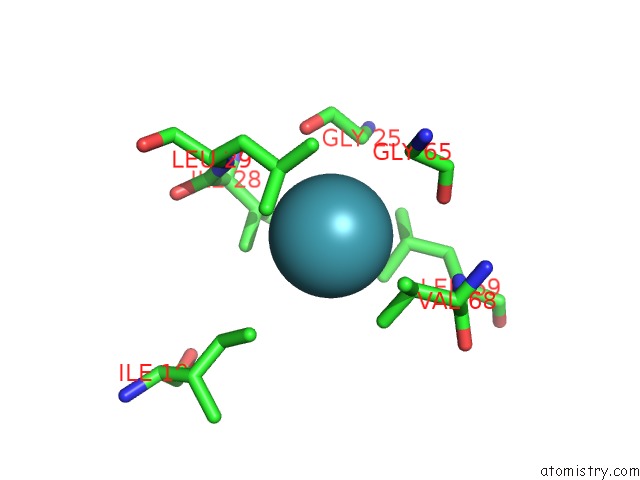
Xenon binding site 1 out of 4 in 2w6w
Go back to
Xenon binding site 1 out
of 4 in the Crystal Structure of Recombinant Sperm Whale Myoglobin Under 1ATM of Xenon
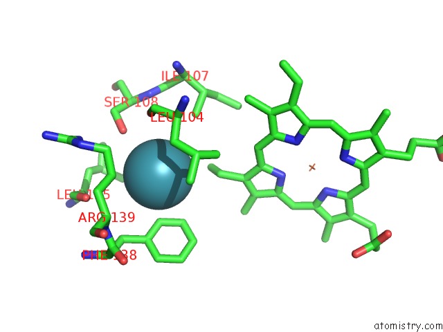
Mono view
Stereo pair view
Mono view
Stereo pair view
A full contact list of Xenon with other atoms in the Xe binding
site number 1 of Crystal Structure of Recombinant Sperm Whale Myoglobin Under 1ATM of Xenon within 5.0Å range:
|
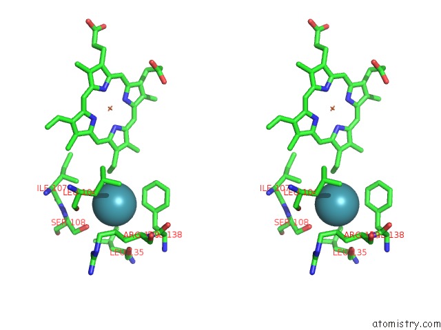
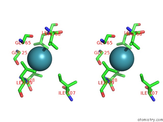
Xenon binding site 2 out of 4 in 2w6w
Go back to
Xenon binding site 2 out
of 4 in the Crystal Structure of Recombinant Sperm Whale Myoglobin Under 1ATM of Xenon
Mono view
Stereo pair view
Mono view
Stereo pair view
A full contact list of Xenon with other atoms in the Xe binding
site number 2 of Crystal Structure of Recombinant Sperm Whale Myoglobin Under 1ATM of Xenon within 5.0Å range:
|
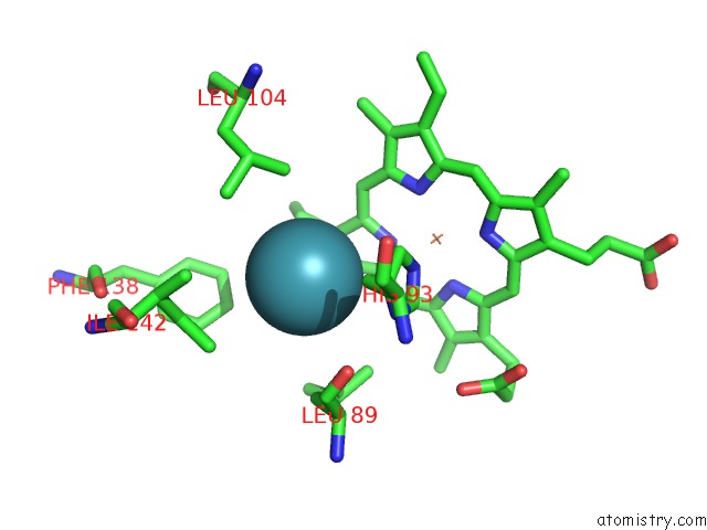
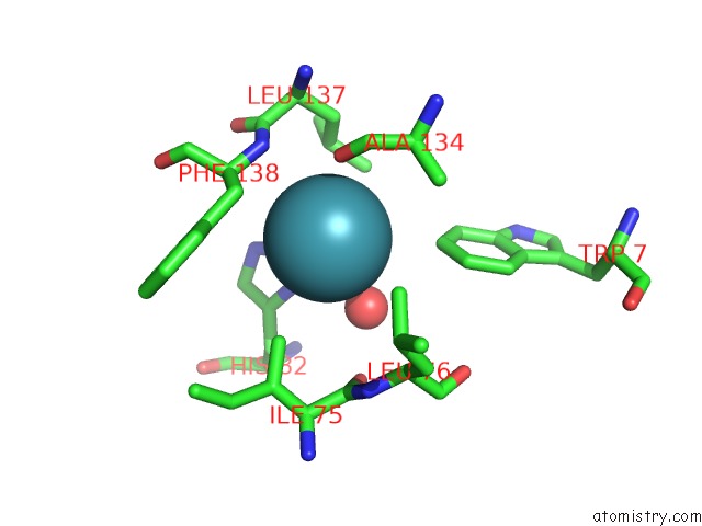
Xenon binding site 3 out of 4 in 2w6w
Go back to
Xenon binding site 3 out
of 4 in the Crystal Structure of Recombinant Sperm Whale Myoglobin Under 1ATM of Xenon
Mono view
Stereo pair view
Mono view
Stereo pair view
A full contact list of Xenon with other atoms in the Xe binding
site number 3 of Crystal Structure of Recombinant Sperm Whale Myoglobin Under 1ATM of Xenon within 5.0Å range:
|
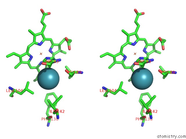
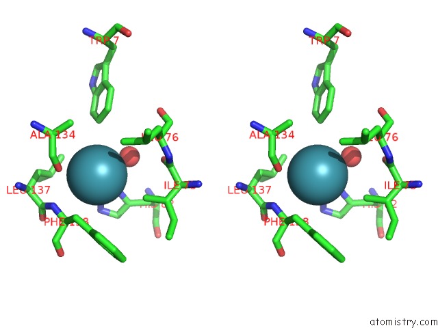
Xenon binding site 4 out of 4 in 2w6w
Go back to
Xenon binding site 4 out
of 4 in the Crystal Structure of Recombinant Sperm Whale Myoglobin Under 1ATM of Xenon
Mono view
Stereo pair view
Mono view
Stereo pair view
A full contact list of Xenon with other atoms in the Xe binding
site number 4 of Crystal Structure of Recombinant Sperm Whale Myoglobin Under 1ATM of Xenon within 5.0Å range:
|
Reference:
C.Savino,
A.E.Miele,
F.Draghi,
K.A.Johnson,
G.Sciara,
M.Brunori,
B.Vallone.
Pattern of Cavities in Globins: the Case of Human Hemoglobin. Biopolymers V. 91 1097 2009.
ISSN: ISSN 0006-3525
PubMed: 19365817
DOI: 10.1002/BIP.21201
Page generated: Sat Oct 12 18:26:34 2024
ISSN: ISSN 0006-3525
PubMed: 19365817
DOI: 10.1002/BIP.21201
Last articles
I in 8D22I in 8CXW
I in 8D00
I in 8CT8
I in 8CUF
I in 8CJ7
I in 8CNZ
I in 8C3P
I in 8CEV
I in 8C7R