Xenon, Xe »
PDB 4zzc-9kum »
5i63 »
Xenon in PDB 5i63: Crystal Structure of TEM1 Beta-Lactamase Mutant I263N in the Presence of 1.2 Mpa Xenon
Enzymatic activity of Crystal Structure of TEM1 Beta-Lactamase Mutant I263N in the Presence of 1.2 Mpa Xenon
All present enzymatic activity of Crystal Structure of TEM1 Beta-Lactamase Mutant I263N in the Presence of 1.2 Mpa Xenon:
3.5.2.6;
3.5.2.6;
Protein crystallography data
The structure of Crystal Structure of TEM1 Beta-Lactamase Mutant I263N in the Presence of 1.2 Mpa Xenon, PDB code: 5i63
was solved by
B.W.Roose,
I.J.Dmochowski,
with X-Ray Crystallography technique. A brief refinement statistics is given in the table below:
| Resolution Low / High (Å) | 60.07 / 1.95 |
| Space group | P 1 21 1 |
| Cell size a, b, c (Å), α, β, γ (°) | 60.070, 84.200, 96.220, 90.00, 89.97, 90.00 |
| R / Rfree (%) | 18.4 / 22.2 |
Xenon Binding Sites:
The binding sites of Xenon atom in the Crystal Structure of TEM1 Beta-Lactamase Mutant I263N in the Presence of 1.2 Mpa Xenon
(pdb code 5i63). This binding sites where shown within
5.0 Angstroms radius around Xenon atom.
In total 8 binding sites of Xenon where determined in the Crystal Structure of TEM1 Beta-Lactamase Mutant I263N in the Presence of 1.2 Mpa Xenon, PDB code: 5i63:
Jump to Xenon binding site number: 1; 2; 3; 4; 5; 6; 7; 8;
In total 8 binding sites of Xenon where determined in the Crystal Structure of TEM1 Beta-Lactamase Mutant I263N in the Presence of 1.2 Mpa Xenon, PDB code: 5i63:
Jump to Xenon binding site number: 1; 2; 3; 4; 5; 6; 7; 8;
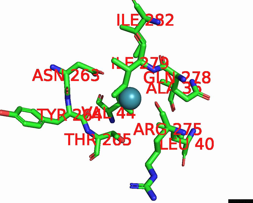
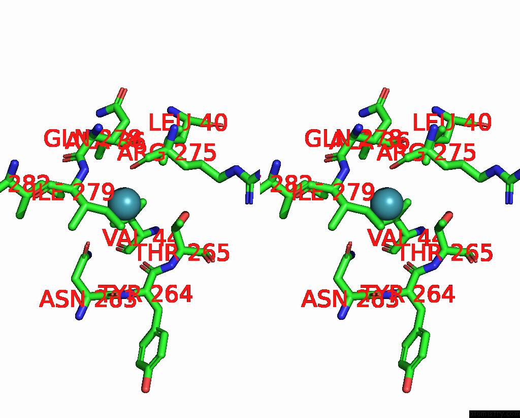
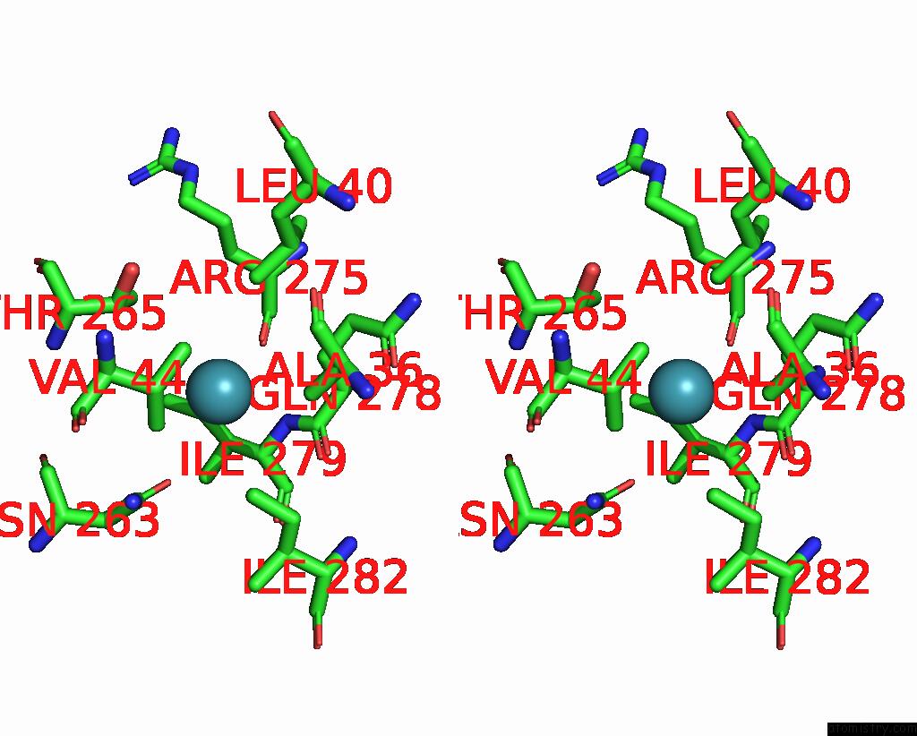
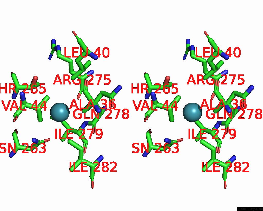
Xenon binding site 1 out of 8 in 5i63
Go back to
Xenon binding site 1 out
of 8 in the Crystal Structure of TEM1 Beta-Lactamase Mutant I263N in the Presence of 1.2 Mpa Xenon
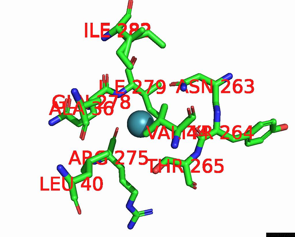
Mono view
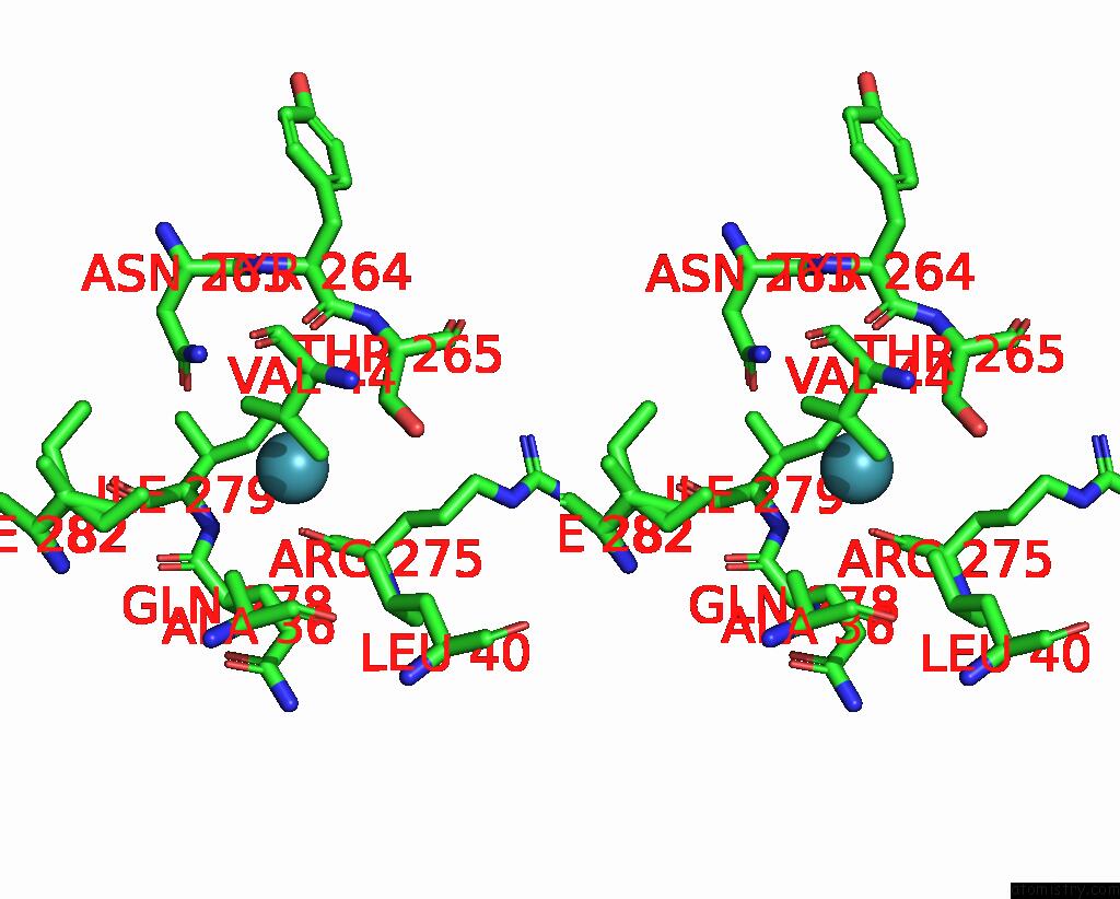
Stereo pair view
Mono view
Stereo pair view
A full contact list of Xenon with other atoms in the Xe binding
site number 1 of Crystal Structure of TEM1 Beta-Lactamase Mutant I263N in the Presence of 1.2 Mpa Xenon within 5.0Å range:
|
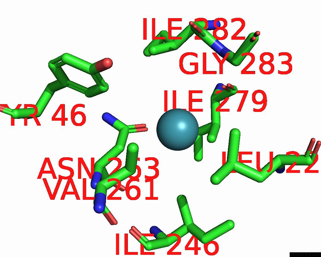
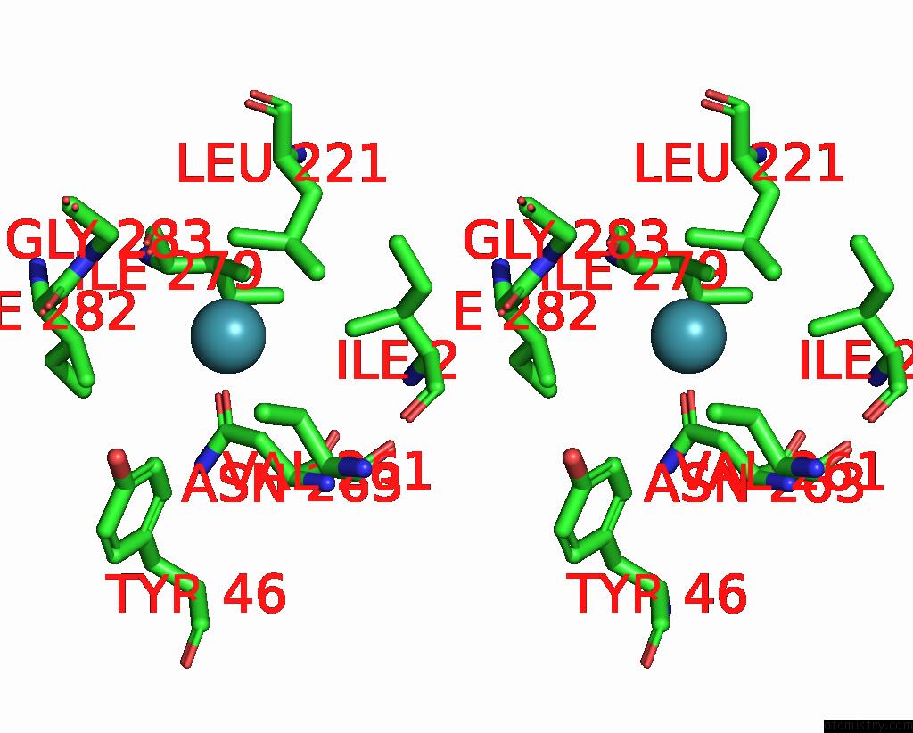
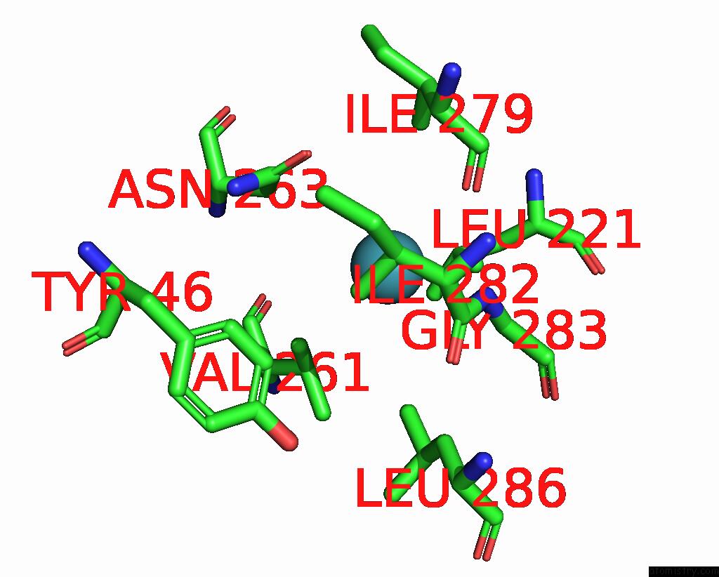
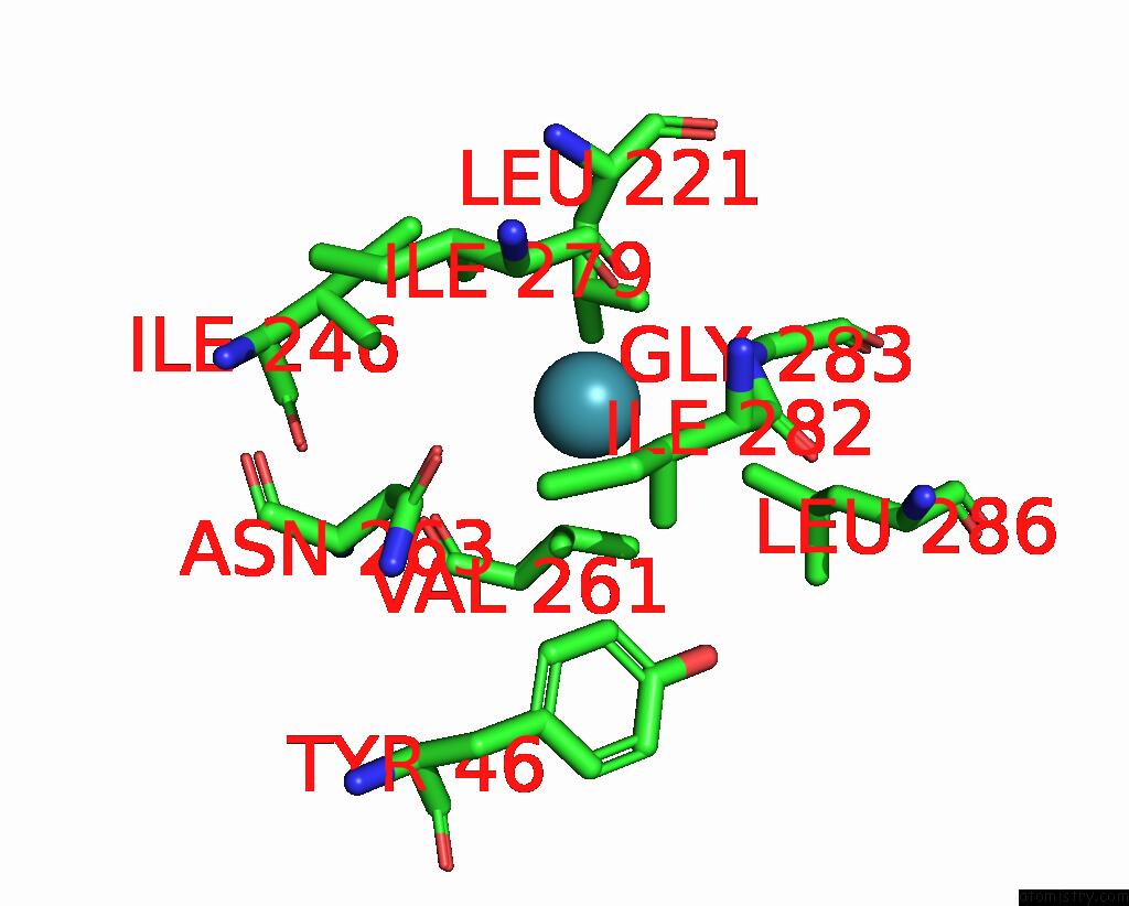
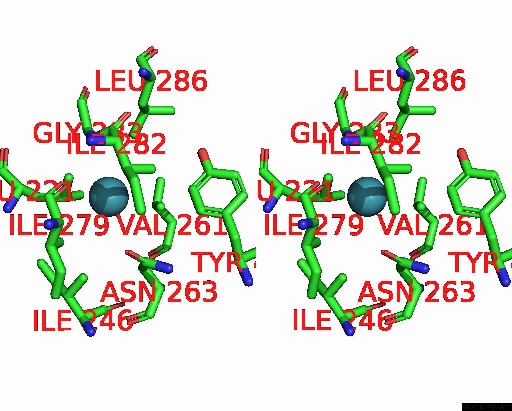
Xenon binding site 2 out of 8 in 5i63
Go back to
Xenon binding site 2 out
of 8 in the Crystal Structure of TEM1 Beta-Lactamase Mutant I263N in the Presence of 1.2 Mpa Xenon
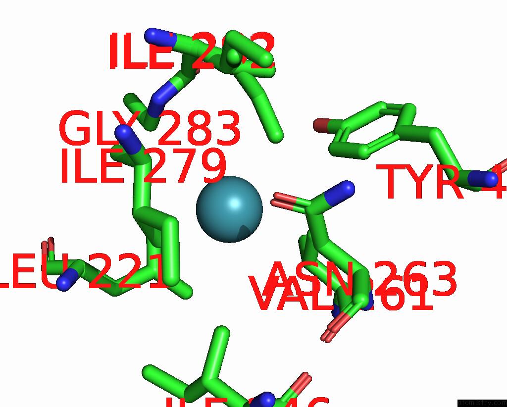
Mono view
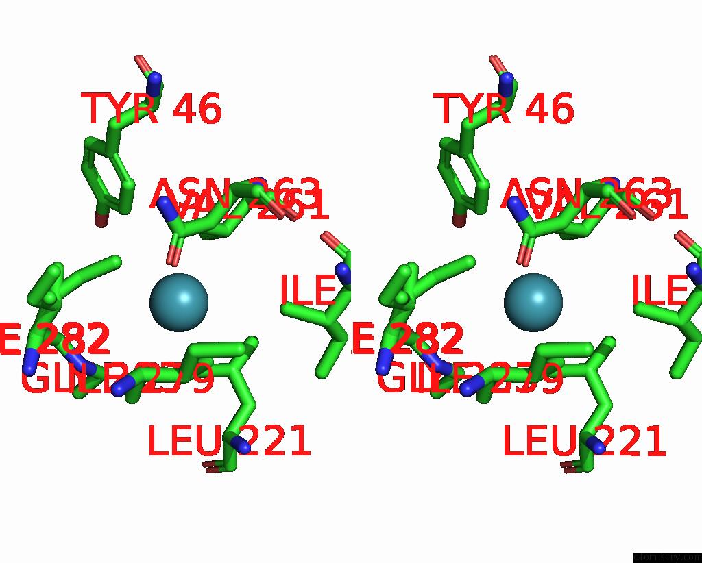
Stereo pair view
Mono view
Stereo pair view
A full contact list of Xenon with other atoms in the Xe binding
site number 2 of Crystal Structure of TEM1 Beta-Lactamase Mutant I263N in the Presence of 1.2 Mpa Xenon within 5.0Å range:
|
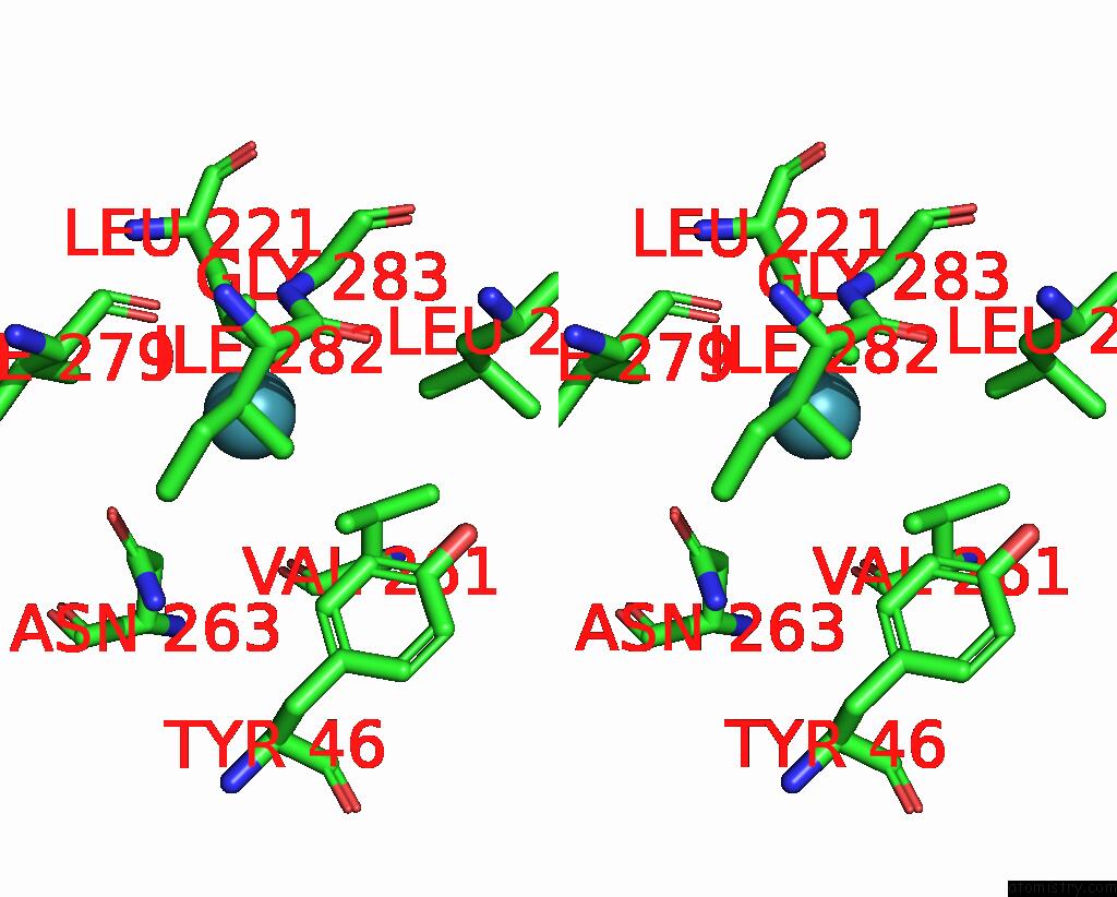
Xenon binding site 3 out of 8 in 5i63
Go back to
Xenon binding site 3 out
of 8 in the Crystal Structure of TEM1 Beta-Lactamase Mutant I263N in the Presence of 1.2 Mpa Xenon
Mono view
Stereo pair view
Mono view
Stereo pair view
A full contact list of Xenon with other atoms in the Xe binding
site number 3 of Crystal Structure of TEM1 Beta-Lactamase Mutant I263N in the Presence of 1.2 Mpa Xenon within 5.0Å range:
|
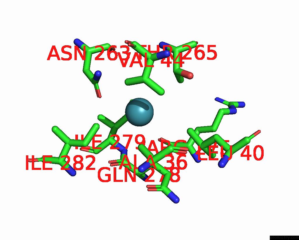
Xenon binding site 4 out of 8 in 5i63
Go back to
Xenon binding site 4 out
of 8 in the Crystal Structure of TEM1 Beta-Lactamase Mutant I263N in the Presence of 1.2 Mpa Xenon
Mono view
Stereo pair view
Mono view
Stereo pair view
A full contact list of Xenon with other atoms in the Xe binding
site number 4 of Crystal Structure of TEM1 Beta-Lactamase Mutant I263N in the Presence of 1.2 Mpa Xenon within 5.0Å range:
|
Xenon binding site 5 out of 8 in 5i63
Go back to
Xenon binding site 5 out
of 8 in the Crystal Structure of TEM1 Beta-Lactamase Mutant I263N in the Presence of 1.2 Mpa Xenon

Mono view
Stereo pair view
Mono view
Stereo pair view
A full contact list of Xenon with other atoms in the Xe binding
site number 5 of Crystal Structure of TEM1 Beta-Lactamase Mutant I263N in the Presence of 1.2 Mpa Xenon within 5.0Å range:
|
Xenon binding site 6 out of 8 in 5i63
Go back to
Xenon binding site 6 out
of 8 in the Crystal Structure of TEM1 Beta-Lactamase Mutant I263N in the Presence of 1.2 Mpa Xenon
Mono view
Stereo pair view
Mono view
Stereo pair view
A full contact list of Xenon with other atoms in the Xe binding
site number 6 of Crystal Structure of TEM1 Beta-Lactamase Mutant I263N in the Presence of 1.2 Mpa Xenon within 5.0Å range:
|
Xenon binding site 7 out of 8 in 5i63
Go back to
Xenon binding site 7 out
of 8 in the Crystal Structure of TEM1 Beta-Lactamase Mutant I263N in the Presence of 1.2 Mpa Xenon
Mono view
Stereo pair view
Mono view
Stereo pair view
A full contact list of Xenon with other atoms in the Xe binding
site number 7 of Crystal Structure of TEM1 Beta-Lactamase Mutant I263N in the Presence of 1.2 Mpa Xenon within 5.0Å range:
|
Xenon binding site 8 out of 8 in 5i63
Go back to
Xenon binding site 8 out
of 8 in the Crystal Structure of TEM1 Beta-Lactamase Mutant I263N in the Presence of 1.2 Mpa Xenon
Mono view
Stereo pair view
Mono view
Stereo pair view
A full contact list of Xenon with other atoms in the Xe binding
site number 8 of Crystal Structure of TEM1 Beta-Lactamase Mutant I263N in the Presence of 1.2 Mpa Xenon within 5.0Å range:
|
Reference:
B.W.Roose,
S.D.Zemerov,
Y.Wang,
M.A.Kasimova,
V.Carnevale,
I.J.Dmochowski.
A Structural Basis FOR129XE Hyper-Cest Signal in Tem-1 Beta-Lactamase. Chemphyschem 2018.
ISSN: ISSN 1439-7641
PubMed: 30151973
DOI: 10.1002/CPHC.201800624
Page generated: Tue Aug 19 18:17:34 2025
ISSN: ISSN 1439-7641
PubMed: 30151973
DOI: 10.1002/CPHC.201800624
Last articles
Zn in 1MKDZn in 1MG0
Zn in 1MKM
Zn in 1ML2
Zn in 1MEY
Zn in 1MGO
Zn in 1MH2
Zn in 1MFS
Zn in 1MFT
Zn in 1MC5