Xenon, Xe »
PDB 3pk5-4zzb »
3tfa »
Xenon in PDB 3tfa: Crystal Structure of An H-Nox Protein From Nostoc Sp. Pcc 7120 Under 6 Atm of Xenon
Protein crystallography data
The structure of Crystal Structure of An H-Nox Protein From Nostoc Sp. Pcc 7120 Under 6 Atm of Xenon, PDB code: 3tfa
was solved by
M.B.Winter,
M.A.Herzik Jr.,
J.Kuriyan,
M.A.Marletta,
with X-Ray Crystallography technique. A brief refinement statistics is given in the table below:
| Resolution Low / High (Å) | 43.82 / 2.27 |
| Space group | P 21 3 |
| Cell size a, b, c (Å), α, β, γ (°) | 123.937, 123.937, 123.937, 90.00, 90.00, 90.00 |
| R / Rfree (%) | 16.3 / 19 |
Other elements in 3tfa:
The structure of Crystal Structure of An H-Nox Protein From Nostoc Sp. Pcc 7120 Under 6 Atm of Xenon also contains other interesting chemical elements:
| Iron | (Fe) | 2 atoms |
Xenon Binding Sites:
The binding sites of Xenon atom in the Crystal Structure of An H-Nox Protein From Nostoc Sp. Pcc 7120 Under 6 Atm of Xenon
(pdb code 3tfa). This binding sites where shown within
5.0 Angstroms radius around Xenon atom.
In total 6 binding sites of Xenon where determined in the Crystal Structure of An H-Nox Protein From Nostoc Sp. Pcc 7120 Under 6 Atm of Xenon, PDB code: 3tfa:
Jump to Xenon binding site number: 1; 2; 3; 4; 5; 6;
In total 6 binding sites of Xenon where determined in the Crystal Structure of An H-Nox Protein From Nostoc Sp. Pcc 7120 Under 6 Atm of Xenon, PDB code: 3tfa:
Jump to Xenon binding site number: 1; 2; 3; 4; 5; 6;
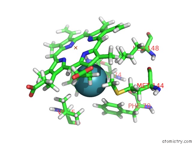
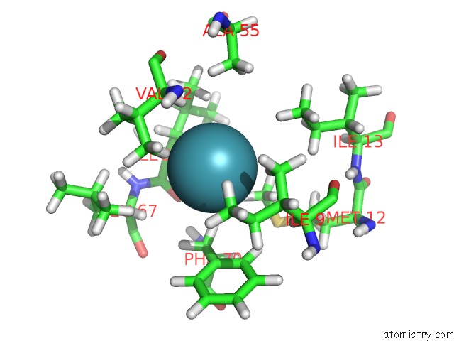
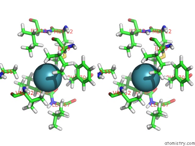
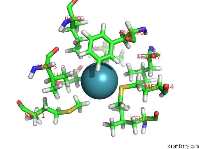
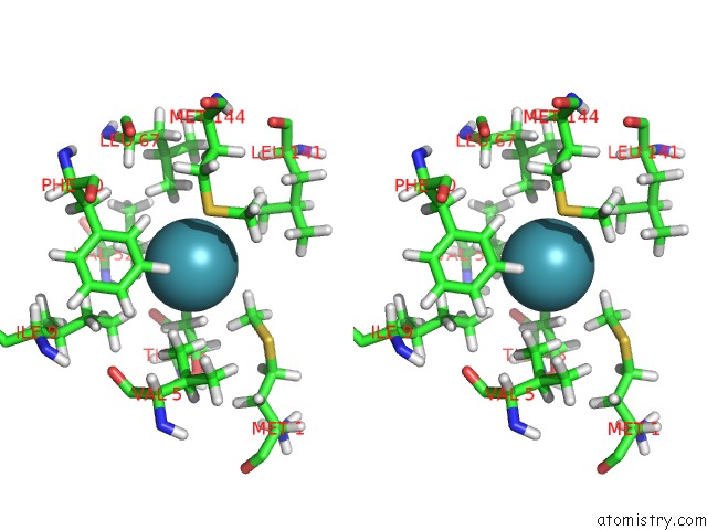
Xenon binding site 1 out of 6 in 3tfa
Go back to
Xenon binding site 1 out
of 6 in the Crystal Structure of An H-Nox Protein From Nostoc Sp. Pcc 7120 Under 6 Atm of Xenon
Mono view
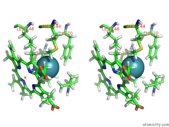
Stereo pair view
Mono view
Stereo pair view
A full contact list of Xenon with other atoms in the Xe binding
site number 1 of Crystal Structure of An H-Nox Protein From Nostoc Sp. Pcc 7120 Under 6 Atm of Xenon within 5.0Å range:
|
Xenon binding site 2 out of 6 in 3tfa
Go back to
Xenon binding site 2 out
of 6 in the Crystal Structure of An H-Nox Protein From Nostoc Sp. Pcc 7120 Under 6 Atm of Xenon
Mono view
Stereo pair view
Mono view
Stereo pair view
A full contact list of Xenon with other atoms in the Xe binding
site number 2 of Crystal Structure of An H-Nox Protein From Nostoc Sp. Pcc 7120 Under 6 Atm of Xenon within 5.0Å range:
|
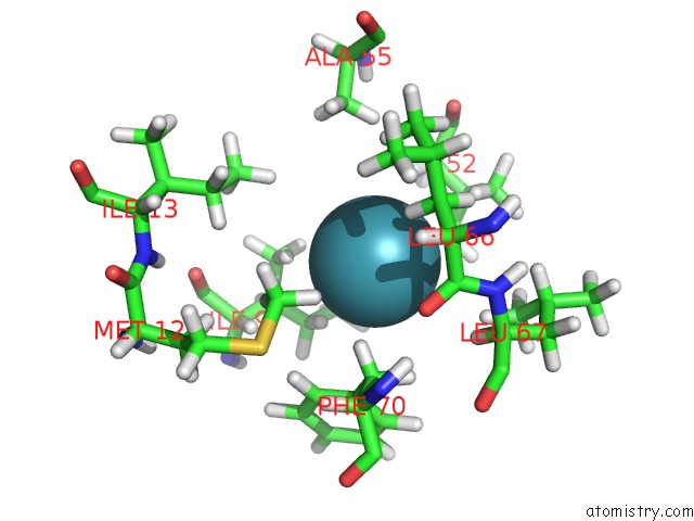
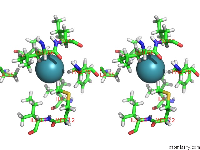
Xenon binding site 3 out of 6 in 3tfa
Go back to
Xenon binding site 3 out
of 6 in the Crystal Structure of An H-Nox Protein From Nostoc Sp. Pcc 7120 Under 6 Atm of Xenon
Mono view
Stereo pair view
Mono view
Stereo pair view
A full contact list of Xenon with other atoms in the Xe binding
site number 3 of Crystal Structure of An H-Nox Protein From Nostoc Sp. Pcc 7120 Under 6 Atm of Xenon within 5.0Å range:
|
Xenon binding site 4 out of 6 in 3tfa
Go back to
Xenon binding site 4 out
of 6 in the Crystal Structure of An H-Nox Protein From Nostoc Sp. Pcc 7120 Under 6 Atm of Xenon
Mono view
Stereo pair view
Mono view
Stereo pair view
A full contact list of Xenon with other atoms in the Xe binding
site number 4 of Crystal Structure of An H-Nox Protein From Nostoc Sp. Pcc 7120 Under 6 Atm of Xenon within 5.0Å range:
|
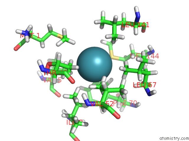
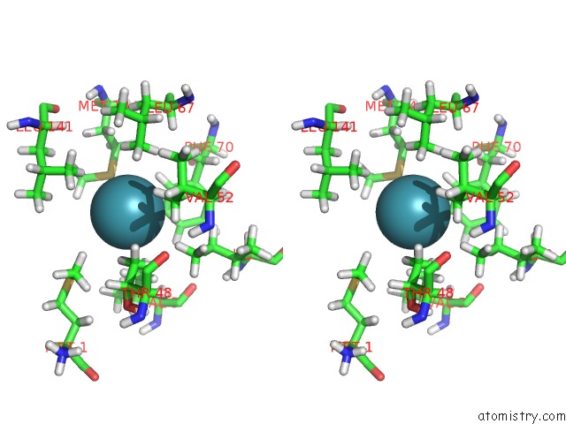
Xenon binding site 5 out of 6 in 3tfa
Go back to
Xenon binding site 5 out
of 6 in the Crystal Structure of An H-Nox Protein From Nostoc Sp. Pcc 7120 Under 6 Atm of Xenon
Mono view
Stereo pair view
Mono view
Stereo pair view
A full contact list of Xenon with other atoms in the Xe binding
site number 5 of Crystal Structure of An H-Nox Protein From Nostoc Sp. Pcc 7120 Under 6 Atm of Xenon within 5.0Å range:
|
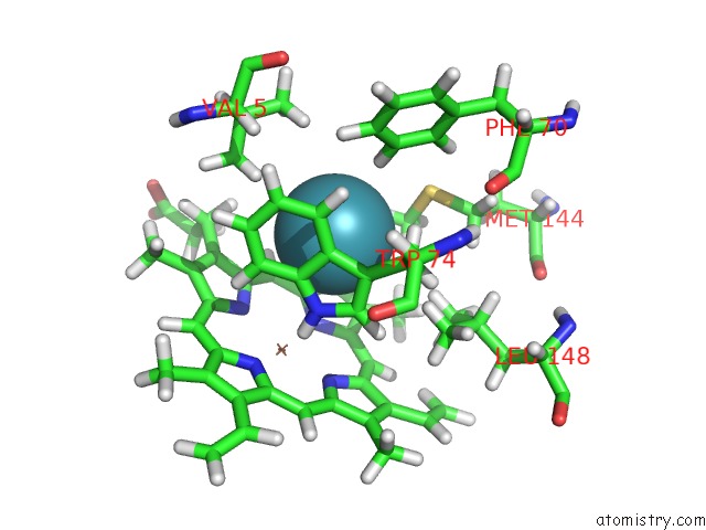
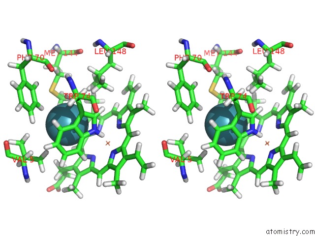
Xenon binding site 6 out of 6 in 3tfa
Go back to
Xenon binding site 6 out
of 6 in the Crystal Structure of An H-Nox Protein From Nostoc Sp. Pcc 7120 Under 6 Atm of Xenon
Mono view
Stereo pair view
Mono view
Stereo pair view
A full contact list of Xenon with other atoms in the Xe binding
site number 6 of Crystal Structure of An H-Nox Protein From Nostoc Sp. Pcc 7120 Under 6 Atm of Xenon within 5.0Å range:
|
Reference:
M.B.Winter,
M.A.Herzik,
J.Kuriyan,
M.A.Marletta.
Tunnels Modulate Ligand Flux in A Heme Nitric Oxide/Oxygen Binding (H-Nox) Domain. Proc.Natl.Acad.Sci.Usa V. 108 E881 2011.
ISSN: ISSN 0027-8424
PubMed: 21997213
DOI: 10.1073/PNAS.1114038108
Page generated: Sat Oct 12 19:06:46 2024
ISSN: ISSN 0027-8424
PubMed: 21997213
DOI: 10.1073/PNAS.1114038108
Last articles
Cl in 5W16Cl in 5W15
Cl in 5W4M
Cl in 5W4I
Cl in 5W4L
Cl in 5W44
Cl in 5W4D
Cl in 5W2O
Cl in 5W2J
Cl in 5VZH