Xenon, Xe »
PDB 1w53-3pk4 »
3g46 »
Xenon in PDB 3g46: Ligand Migration and Cavities Within Scapharca Dimeric Hemoglobin: Wild Type with Co Bound to Heme and Chloroform Bound to the XE4 Cavity
Protein crystallography data
The structure of Ligand Migration and Cavities Within Scapharca Dimeric Hemoglobin: Wild Type with Co Bound to Heme and Chloroform Bound to the XE4 Cavity, PDB code: 3g46
was solved by
J.E.Knapp,
R.Pahl,
J.Cohen,
J.C.Nichols,
K.Schulten,
Q.H.Gibson,
V.Srajer,
W.E.Royer Jr.,
with X-Ray Crystallography technique. A brief refinement statistics is given in the table below:
| Resolution Low / High (Å) | 46.30 / 0.91 |
| Space group | C 1 2 1 |
| Cell size a, b, c (Å), α, β, γ (°) | 92.536, 43.487, 82.853, 90.00, 122.25, 90.00 |
| R / Rfree (%) | 12.9 / 15.6 |
Other elements in 3g46:
The structure of Ligand Migration and Cavities Within Scapharca Dimeric Hemoglobin: Wild Type with Co Bound to Heme and Chloroform Bound to the XE4 Cavity also contains other interesting chemical elements:
| Iron | (Fe) | 2 atoms |
Xenon Binding Sites:
The binding sites of Xenon atom in the Ligand Migration and Cavities Within Scapharca Dimeric Hemoglobin: Wild Type with Co Bound to Heme and Chloroform Bound to the XE4 Cavity
(pdb code 3g46). This binding sites where shown within
5.0 Angstroms radius around Xenon atom.
In total 8 binding sites of Xenon where determined in the Ligand Migration and Cavities Within Scapharca Dimeric Hemoglobin: Wild Type with Co Bound to Heme and Chloroform Bound to the XE4 Cavity, PDB code: 3g46:
Jump to Xenon binding site number: 1; 2; 3; 4; 5; 6; 7; 8;
In total 8 binding sites of Xenon where determined in the Ligand Migration and Cavities Within Scapharca Dimeric Hemoglobin: Wild Type with Co Bound to Heme and Chloroform Bound to the XE4 Cavity, PDB code: 3g46:
Jump to Xenon binding site number: 1; 2; 3; 4; 5; 6; 7; 8;
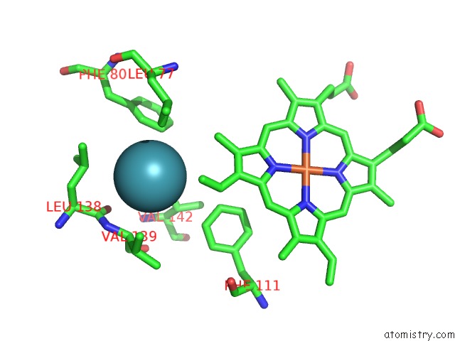
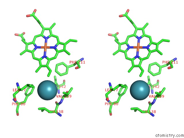
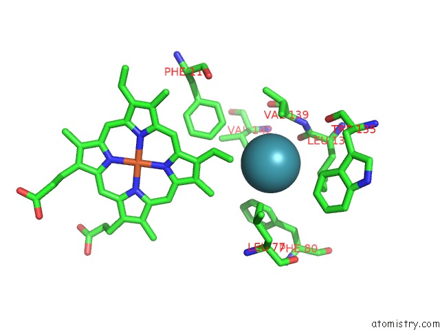
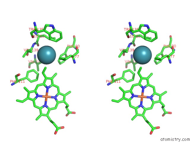
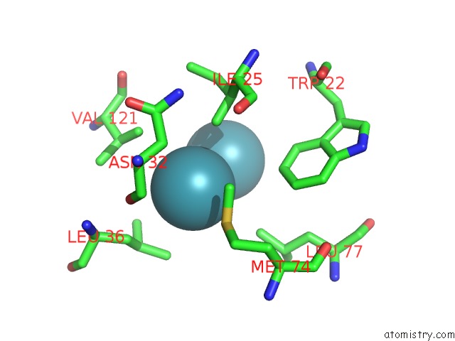
Xenon binding site 1 out of 8 in 3g46
Go back to
Xenon binding site 1 out
of 8 in the Ligand Migration and Cavities Within Scapharca Dimeric Hemoglobin: Wild Type with Co Bound to Heme and Chloroform Bound to the XE4 Cavity
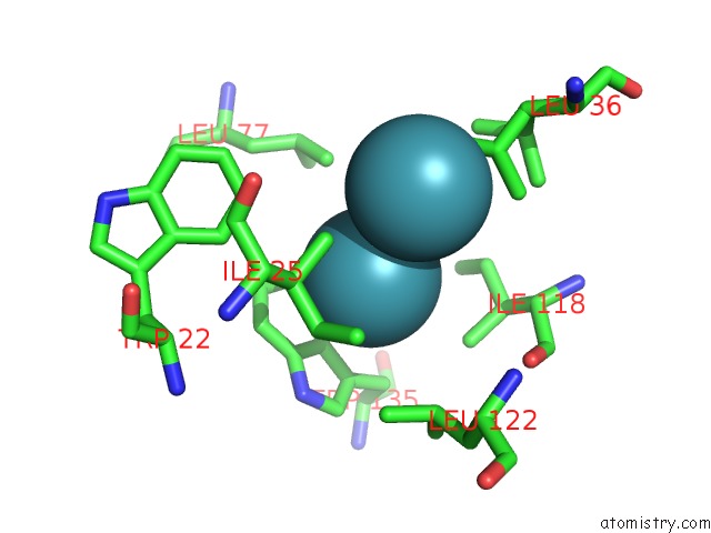
Mono view
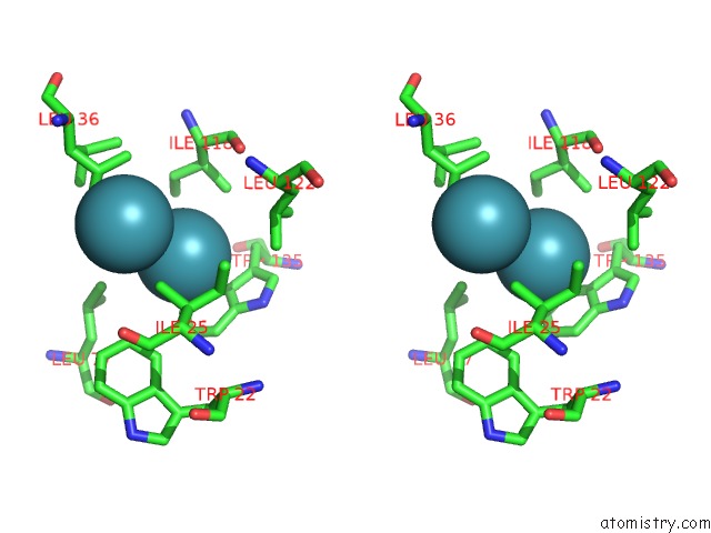
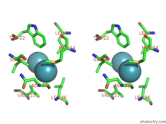
Stereo pair view
Mono view
Stereo pair view
A full contact list of Xenon with other atoms in the Xe binding
site number 1 of Ligand Migration and Cavities Within Scapharca Dimeric Hemoglobin: Wild Type with Co Bound to Heme and Chloroform Bound to the XE4 Cavity within 5.0Å range:
|
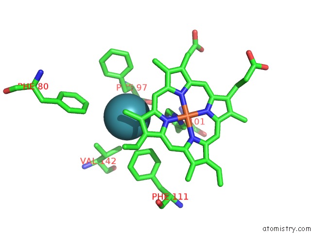
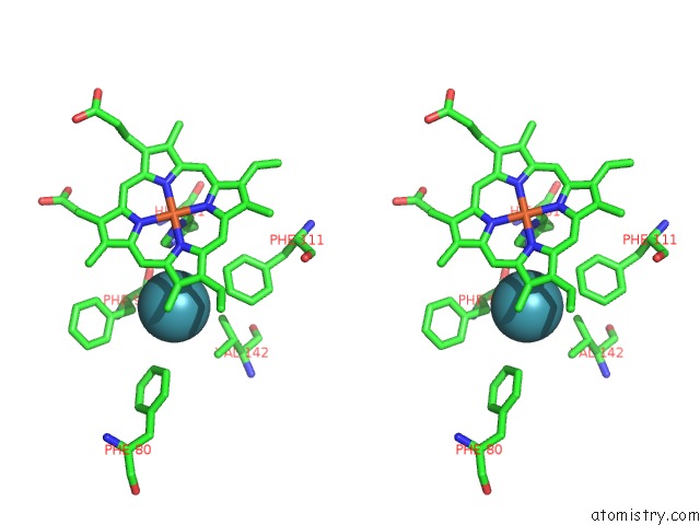
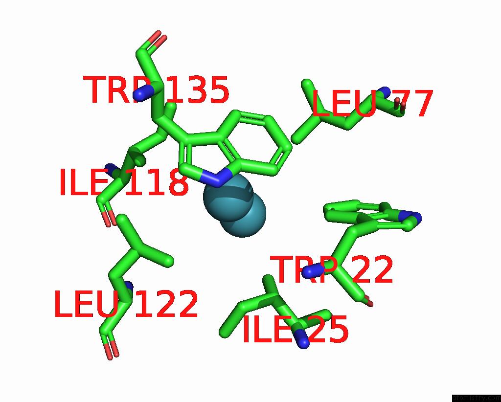
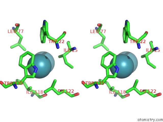
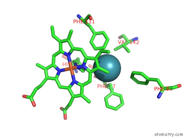
Xenon binding site 2 out of 8 in 3g46
Go back to
Xenon binding site 2 out
of 8 in the Ligand Migration and Cavities Within Scapharca Dimeric Hemoglobin: Wild Type with Co Bound to Heme and Chloroform Bound to the XE4 Cavity
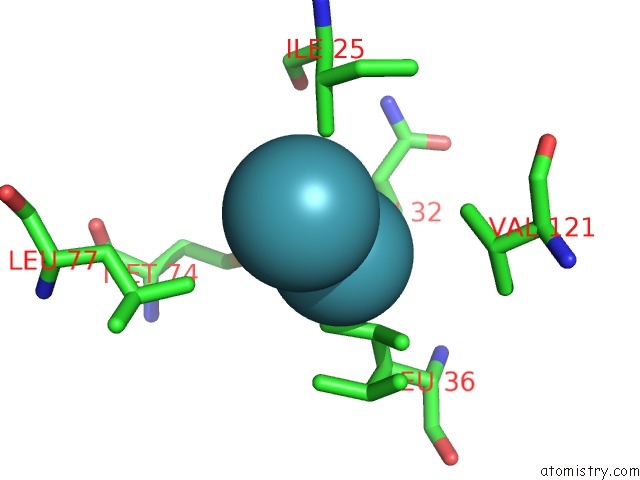
Mono view
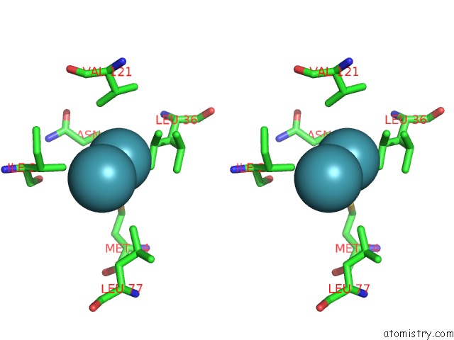
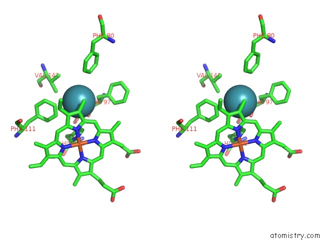
Stereo pair view
Mono view
Stereo pair view
A full contact list of Xenon with other atoms in the Xe binding
site number 2 of Ligand Migration and Cavities Within Scapharca Dimeric Hemoglobin: Wild Type with Co Bound to Heme and Chloroform Bound to the XE4 Cavity within 5.0Å range:
|
Xenon binding site 3 out of 8 in 3g46
Go back to
Xenon binding site 3 out
of 8 in the Ligand Migration and Cavities Within Scapharca Dimeric Hemoglobin: Wild Type with Co Bound to Heme and Chloroform Bound to the XE4 Cavity
Mono view
Stereo pair view
Mono view
Stereo pair view
A full contact list of Xenon with other atoms in the Xe binding
site number 3 of Ligand Migration and Cavities Within Scapharca Dimeric Hemoglobin: Wild Type with Co Bound to Heme and Chloroform Bound to the XE4 Cavity within 5.0Å range:
|
Xenon binding site 4 out of 8 in 3g46
Go back to
Xenon binding site 4 out
of 8 in the Ligand Migration and Cavities Within Scapharca Dimeric Hemoglobin: Wild Type with Co Bound to Heme and Chloroform Bound to the XE4 Cavity
Mono view
Stereo pair view
Mono view
Stereo pair view
A full contact list of Xenon with other atoms in the Xe binding
site number 4 of Ligand Migration and Cavities Within Scapharca Dimeric Hemoglobin: Wild Type with Co Bound to Heme and Chloroform Bound to the XE4 Cavity within 5.0Å range:
|
Xenon binding site 5 out of 8 in 3g46
Go back to
Xenon binding site 5 out
of 8 in the Ligand Migration and Cavities Within Scapharca Dimeric Hemoglobin: Wild Type with Co Bound to Heme and Chloroform Bound to the XE4 Cavity
Mono view
Stereo pair view
Mono view
Stereo pair view
A full contact list of Xenon with other atoms in the Xe binding
site number 5 of Ligand Migration and Cavities Within Scapharca Dimeric Hemoglobin: Wild Type with Co Bound to Heme and Chloroform Bound to the XE4 Cavity within 5.0Å range:
|
Xenon binding site 6 out of 8 in 3g46
Go back to
Xenon binding site 6 out
of 8 in the Ligand Migration and Cavities Within Scapharca Dimeric Hemoglobin: Wild Type with Co Bound to Heme and Chloroform Bound to the XE4 Cavity
Mono view
Stereo pair view
Mono view
Stereo pair view
A full contact list of Xenon with other atoms in the Xe binding
site number 6 of Ligand Migration and Cavities Within Scapharca Dimeric Hemoglobin: Wild Type with Co Bound to Heme and Chloroform Bound to the XE4 Cavity within 5.0Å range:
|
Xenon binding site 7 out of 8 in 3g46
Go back to
Xenon binding site 7 out
of 8 in the Ligand Migration and Cavities Within Scapharca Dimeric Hemoglobin: Wild Type with Co Bound to Heme and Chloroform Bound to the XE4 Cavity
Mono view
Stereo pair view
Mono view
Stereo pair view
A full contact list of Xenon with other atoms in the Xe binding
site number 7 of Ligand Migration and Cavities Within Scapharca Dimeric Hemoglobin: Wild Type with Co Bound to Heme and Chloroform Bound to the XE4 Cavity within 5.0Å range:
|
Xenon binding site 8 out of 8 in 3g46
Go back to
Xenon binding site 8 out
of 8 in the Ligand Migration and Cavities Within Scapharca Dimeric Hemoglobin: Wild Type with Co Bound to Heme and Chloroform Bound to the XE4 Cavity
Mono view
Stereo pair view
Mono view
Stereo pair view
A full contact list of Xenon with other atoms in the Xe binding
site number 8 of Ligand Migration and Cavities Within Scapharca Dimeric Hemoglobin: Wild Type with Co Bound to Heme and Chloroform Bound to the XE4 Cavity within 5.0Å range:
|
Reference:
J.E.Knapp,
R.Pahl,
J.Cohen,
J.C.Nichols,
K.Schulten,
Q.H.Gibson,
V.Srajer,
W.E.Royer.
Ligand Migration and Cavities Within Scapharca Dimeric Hbi: Studies By Time-Resolved Crystallo-Graphy, Xe Binding, and Computational Analysis. Structure V. 17 1494 2009.
ISSN: ISSN 0969-2126
PubMed: 19913484
DOI: 10.1016/J.STR.2009.09.004
Page generated: Sat Oct 12 18:46:16 2024
ISSN: ISSN 0969-2126
PubMed: 19913484
DOI: 10.1016/J.STR.2009.09.004
Last articles
Zn in 9MJ5Zn in 9HNW
Zn in 9G0L
Zn in 9FNE
Zn in 9DZN
Zn in 9E0I
Zn in 9D32
Zn in 9DAK
Zn in 8ZXC
Zn in 8ZUF