Xenon, Xe »
PDB 1w53-3pk4 »
2w72 »
Xenon in PDB 2w72: Deoxygenated Structure of A Distal Site Hemoglobin Mutant Plus Xe
Protein crystallography data
The structure of Deoxygenated Structure of A Distal Site Hemoglobin Mutant Plus Xe, PDB code: 2w72
was solved by
A.E.Miele,
F.Draghi,
G.Sciara,
K.A.Johnson,
F.Renzi,
B.Vallone,
M.Brunori,
C.Savino,
with X-Ray Crystallography technique. A brief refinement statistics is given in the table below:
| Resolution Low / High (Å) | 17.32 / 1.07 |
| Space group | P 1 21 1 |
| Cell size a, b, c (Å), α, β, γ (°) | 61.957, 82.447, 53.531, 90.00, 99.60, 90.00 |
| R / Rfree (%) | 12.9 / 15.3 |
Other elements in 2w72:
The structure of Deoxygenated Structure of A Distal Site Hemoglobin Mutant Plus Xe also contains other interesting chemical elements:
| Potassium | (K) | 3 atoms |
| Iron | (Fe) | 4 atoms |
Xenon Binding Sites:
Pages:
>>> Page 1 <<< Page 2, Binding sites: 11 - 20; Page 3, Binding sites: 21 - 23;Binding sites:
The binding sites of Xenon atom in the Deoxygenated Structure of A Distal Site Hemoglobin Mutant Plus Xe (pdb code 2w72). This binding sites where shown within 5.0 Angstroms radius around Xenon atom.In total 23 binding sites of Xenon where determined in the Deoxygenated Structure of A Distal Site Hemoglobin Mutant Plus Xe, PDB code: 2w72:
Jump to Xenon binding site number: 1; 2; 3; 4; 5; 6; 7; 8; 9; 10;
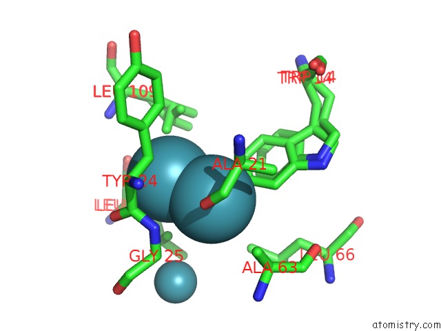
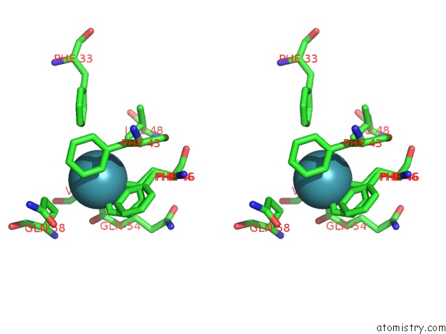
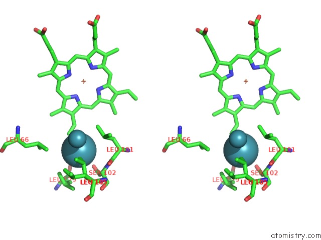
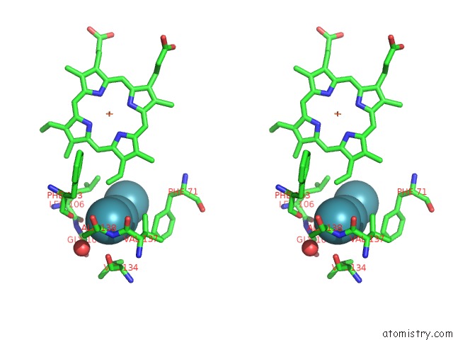
Xenon binding site 1 out of 23 in 2w72
Go back to
Xenon binding site 1 out
of 23 in the Deoxygenated Structure of A Distal Site Hemoglobin Mutant Plus Xe
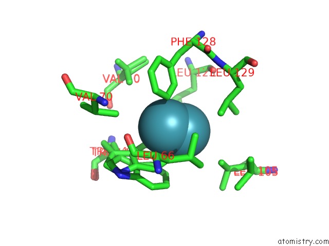
Mono view
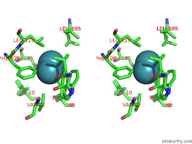
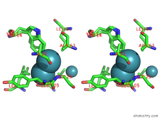
Stereo pair view
Mono view
Stereo pair view
A full contact list of Xenon with other atoms in the Xe binding
site number 1 of Deoxygenated Structure of A Distal Site Hemoglobin Mutant Plus Xe within 5.0Å range:
|
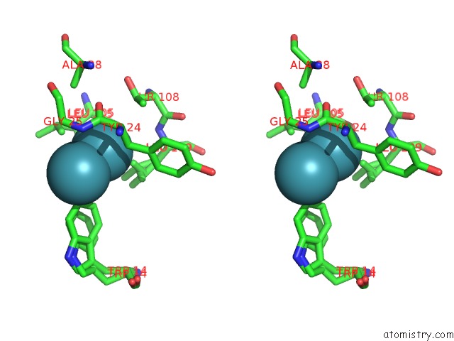
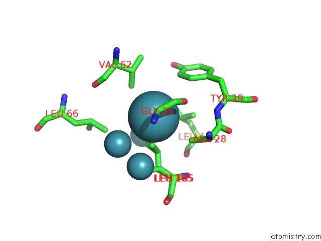
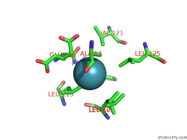
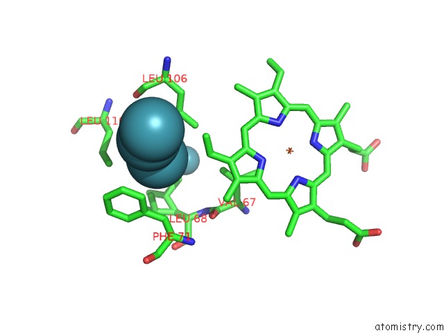
Xenon binding site 2 out of 23 in 2w72
Go back to
Xenon binding site 2 out
of 23 in the Deoxygenated Structure of A Distal Site Hemoglobin Mutant Plus Xe
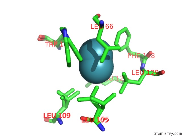
Mono view
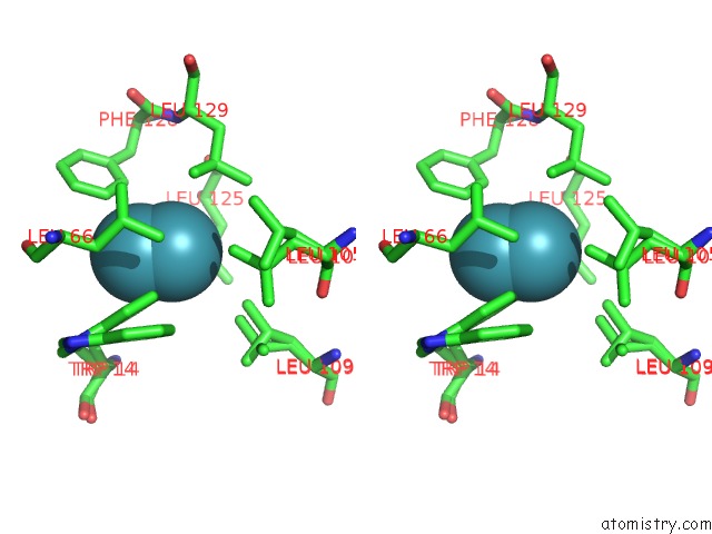
Stereo pair view
Mono view
Stereo pair view
A full contact list of Xenon with other atoms in the Xe binding
site number 2 of Deoxygenated Structure of A Distal Site Hemoglobin Mutant Plus Xe within 5.0Å range:
|
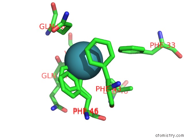
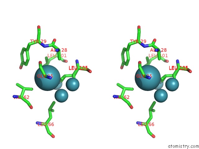
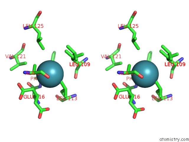
Xenon binding site 3 out of 23 in 2w72
Go back to
Xenon binding site 3 out
of 23 in the Deoxygenated Structure of A Distal Site Hemoglobin Mutant Plus Xe
Mono view
Stereo pair view
Mono view
Stereo pair view
A full contact list of Xenon with other atoms in the Xe binding
site number 3 of Deoxygenated Structure of A Distal Site Hemoglobin Mutant Plus Xe within 5.0Å range:
|
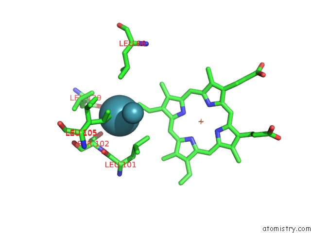
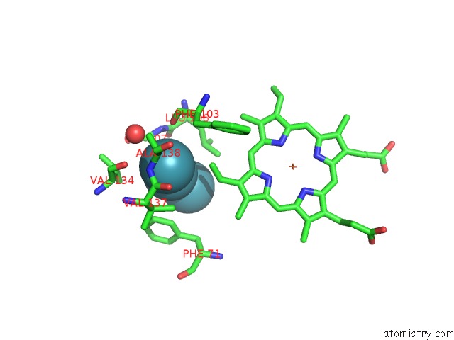
Xenon binding site 4 out of 23 in 2w72
Go back to
Xenon binding site 4 out
of 23 in the Deoxygenated Structure of A Distal Site Hemoglobin Mutant Plus Xe

Mono view
Stereo pair view
Mono view
Stereo pair view
A full contact list of Xenon with other atoms in the Xe binding
site number 4 of Deoxygenated Structure of A Distal Site Hemoglobin Mutant Plus Xe within 5.0Å range:
|
Xenon binding site 5 out of 23 in 2w72
Go back to
Xenon binding site 5 out
of 23 in the Deoxygenated Structure of A Distal Site Hemoglobin Mutant Plus Xe
Mono view
Stereo pair view
Mono view
Stereo pair view
A full contact list of Xenon with other atoms in the Xe binding
site number 5 of Deoxygenated Structure of A Distal Site Hemoglobin Mutant Plus Xe within 5.0Å range:
|
Xenon binding site 6 out of 23 in 2w72
Go back to
Xenon binding site 6 out
of 23 in the Deoxygenated Structure of A Distal Site Hemoglobin Mutant Plus Xe
Mono view
Stereo pair view
Mono view
Stereo pair view
A full contact list of Xenon with other atoms in the Xe binding
site number 6 of Deoxygenated Structure of A Distal Site Hemoglobin Mutant Plus Xe within 5.0Å range:
|
Xenon binding site 7 out of 23 in 2w72
Go back to
Xenon binding site 7 out
of 23 in the Deoxygenated Structure of A Distal Site Hemoglobin Mutant Plus Xe
Mono view
Stereo pair view
Mono view
Stereo pair view
A full contact list of Xenon with other atoms in the Xe binding
site number 7 of Deoxygenated Structure of A Distal Site Hemoglobin Mutant Plus Xe within 5.0Å range:
|
Xenon binding site 8 out of 23 in 2w72
Go back to
Xenon binding site 8 out
of 23 in the Deoxygenated Structure of A Distal Site Hemoglobin Mutant Plus Xe
Mono view
Stereo pair view
Mono view
Stereo pair view
A full contact list of Xenon with other atoms in the Xe binding
site number 8 of Deoxygenated Structure of A Distal Site Hemoglobin Mutant Plus Xe within 5.0Å range:
|
Xenon binding site 9 out of 23 in 2w72
Go back to
Xenon binding site 9 out
of 23 in the Deoxygenated Structure of A Distal Site Hemoglobin Mutant Plus Xe
Mono view
Stereo pair view
Mono view
Stereo pair view
A full contact list of Xenon with other atoms in the Xe binding
site number 9 of Deoxygenated Structure of A Distal Site Hemoglobin Mutant Plus Xe within 5.0Å range:
|
Xenon binding site 10 out of 23 in 2w72
Go back to
Xenon binding site 10 out
of 23 in the Deoxygenated Structure of A Distal Site Hemoglobin Mutant Plus Xe
Mono view

Stereo pair view
Mono view
Stereo pair view
A full contact list of Xenon with other atoms in the Xe binding
site number 10 of Deoxygenated Structure of A Distal Site Hemoglobin Mutant Plus Xe within 5.0Å range:
|
Reference:
C.Savino,
A.E.Miele,
F.Draghi,
K.A.Johnson,
G.Sciara,
M.Brunori,
B.Vallone.
Pattern of Cavities in Globins: the Case of Human Hemoglobin. Biopolymers V. 91 1097 2009.
ISSN: ISSN 0006-3525
PubMed: 19365817
DOI: 10.1002/BIP.21201
Page generated: Sat Oct 12 18:29:25 2024
ISSN: ISSN 0006-3525
PubMed: 19365817
DOI: 10.1002/BIP.21201
Last articles
Cl in 4ZJWCl in 4ZJJ
Cl in 4ZJI
Cl in 4ZJM
Cl in 4ZIX
Cl in 4ZJ8
Cl in 4ZIM
Cl in 4ZIR
Cl in 4ZIQ
Cl in 4ZIP