Xenon, Xe »
PDB 1w53-3pk4 »
2w6y »
Xenon in PDB 2w6y: Crystal Structure of Sperm Whale Myoglobin Mutant Yqr in Complex with Xenon
Protein crystallography data
The structure of Crystal Structure of Sperm Whale Myoglobin Mutant Yqr in Complex with Xenon, PDB code: 2w6y
was solved by
A.E.Miele,
F.Draghi,
F.Renzi,
G.Sciara,
K.A.Johnson,
B.Vallone,
M.Brunori,
C.Savino,
with X-Ray Crystallography technique. A brief refinement statistics is given in the table below:
| Resolution Low / High (Å) | 26.00 / 1.60 |
| Space group | P 6 |
| Cell size a, b, c (Å), α, β, γ (°) | 90.626, 90.626, 45.505, 90.00, 90.00, 120.00 |
| R / Rfree (%) | 16.5 / 19.6 |
Other elements in 2w6y:
The structure of Crystal Structure of Sperm Whale Myoglobin Mutant Yqr in Complex with Xenon also contains other interesting chemical elements:
| Iron | (Fe) | 1 atom |
Xenon Binding Sites:
The binding sites of Xenon atom in the Crystal Structure of Sperm Whale Myoglobin Mutant Yqr in Complex with Xenon
(pdb code 2w6y). This binding sites where shown within
5.0 Angstroms radius around Xenon atom.
In total 4 binding sites of Xenon where determined in the Crystal Structure of Sperm Whale Myoglobin Mutant Yqr in Complex with Xenon, PDB code: 2w6y:
Jump to Xenon binding site number: 1; 2; 3; 4;
In total 4 binding sites of Xenon where determined in the Crystal Structure of Sperm Whale Myoglobin Mutant Yqr in Complex with Xenon, PDB code: 2w6y:
Jump to Xenon binding site number: 1; 2; 3; 4;
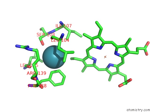
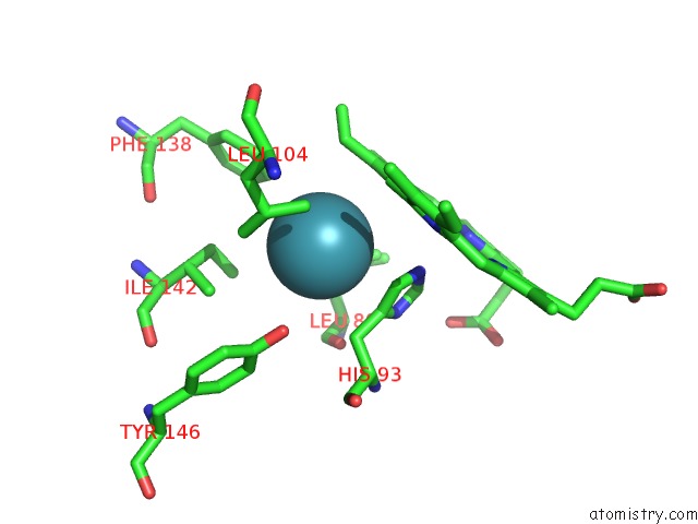
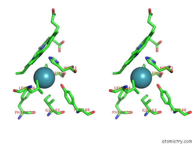
Xenon binding site 1 out of 4 in 2w6y
Go back to
Xenon binding site 1 out
of 4 in the Crystal Structure of Sperm Whale Myoglobin Mutant Yqr in Complex with Xenon
Mono view
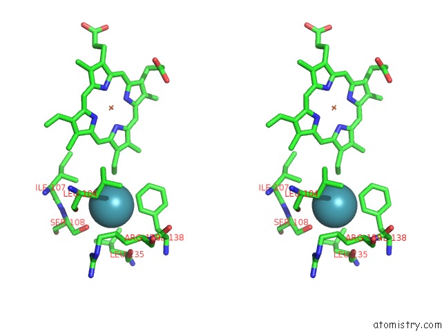
Stereo pair view
Mono view
Stereo pair view
A full contact list of Xenon with other atoms in the Xe binding
site number 1 of Crystal Structure of Sperm Whale Myoglobin Mutant Yqr in Complex with Xenon within 5.0Å range:
|
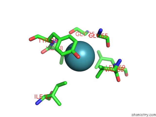
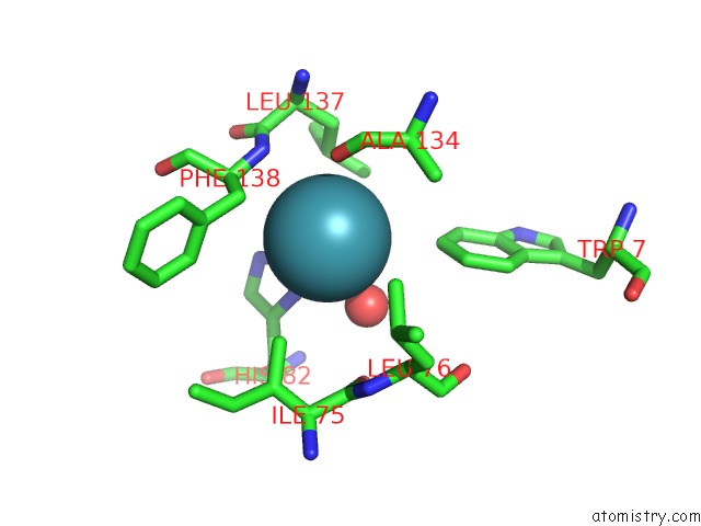
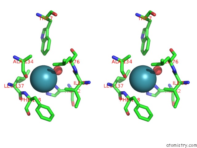
Xenon binding site 2 out of 4 in 2w6y
Go back to
Xenon binding site 2 out
of 4 in the Crystal Structure of Sperm Whale Myoglobin Mutant Yqr in Complex with Xenon
Mono view
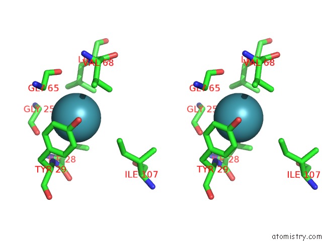
Stereo pair view
Mono view
Stereo pair view
A full contact list of Xenon with other atoms in the Xe binding
site number 2 of Crystal Structure of Sperm Whale Myoglobin Mutant Yqr in Complex with Xenon within 5.0Å range:
|
Xenon binding site 3 out of 4 in 2w6y
Go back to
Xenon binding site 3 out
of 4 in the Crystal Structure of Sperm Whale Myoglobin Mutant Yqr in Complex with Xenon
Mono view
Stereo pair view
Mono view
Stereo pair view
A full contact list of Xenon with other atoms in the Xe binding
site number 3 of Crystal Structure of Sperm Whale Myoglobin Mutant Yqr in Complex with Xenon within 5.0Å range:
|
Xenon binding site 4 out of 4 in 2w6y
Go back to
Xenon binding site 4 out
of 4 in the Crystal Structure of Sperm Whale Myoglobin Mutant Yqr in Complex with Xenon
Mono view
Stereo pair view
Mono view
Stereo pair view
A full contact list of Xenon with other atoms in the Xe binding
site number 4 of Crystal Structure of Sperm Whale Myoglobin Mutant Yqr in Complex with Xenon within 5.0Å range:
|
Reference:
A.E.Miele,
F.Draghi,
K.A.Johnson,
F.Renzi,
G.Sciara,
M.Brunori,
B.Vallone,
C.Savino.
When the Same Fold Does Not Mean the Same Function: the Case of Xenon Cavities in Hemoglobin and Myoglobin To Be Published.
Page generated: Tue Aug 19 17:54:06 2025
Last articles
Zn in 1MWQZn in 1MWO
Zn in 1MXG
Zn in 1MWZ
Zn in 1MS7
Zn in 1MVX
Zn in 1MVH
Zn in 1MUA
Zn in 1MSO
Zn in 1MRO