Xenon, Xe »
PDB 1w53-3pk4 »
2a7c »
Xenon in PDB 2a7c: On the Routine Use of Soft X-Rays in Macromolecular Crystallography, Part III- the Optimal Data Collection Wavelength
Enzymatic activity of On the Routine Use of Soft X-Rays in Macromolecular Crystallography, Part III- the Optimal Data Collection Wavelength
All present enzymatic activity of On the Routine Use of Soft X-Rays in Macromolecular Crystallography, Part III- the Optimal Data Collection Wavelength:
3.4.21.36;
3.4.21.36;
Protein crystallography data
The structure of On the Routine Use of Soft X-Rays in Macromolecular Crystallography, Part III- the Optimal Data Collection Wavelength, PDB code: 2a7c
was solved by
C.Mueller-Dieckmann,
S.Panjikar,
P.A.Tucker,
M.S.Weiss,
with X-Ray Crystallography technique. A brief refinement statistics is given in the table below:
| Resolution Low / High (Å) | 40.00 / 1.65 |
| Space group | P 21 21 21 |
| Cell size a, b, c (Å), α, β, γ (°) | 50.350, 58.020, 74.740, 90.00, 90.00, 90.00 |
| R / Rfree (%) | 15.5 / 18.2 |
Xenon Binding Sites:
The binding sites of Xenon atom in the On the Routine Use of Soft X-Rays in Macromolecular Crystallography, Part III- the Optimal Data Collection Wavelength
(pdb code 2a7c). This binding sites where shown within
5.0 Angstroms radius around Xenon atom.
In total only one binding site of Xenon was determined in the On the Routine Use of Soft X-Rays in Macromolecular Crystallography, Part III- the Optimal Data Collection Wavelength, PDB code: 2a7c:
In total only one binding site of Xenon was determined in the On the Routine Use of Soft X-Rays in Macromolecular Crystallography, Part III- the Optimal Data Collection Wavelength, PDB code: 2a7c:
Xenon binding site 1 out of 1 in 2a7c
Go back to
Xenon binding site 1 out
of 1 in the On the Routine Use of Soft X-Rays in Macromolecular Crystallography, Part III- the Optimal Data Collection Wavelength
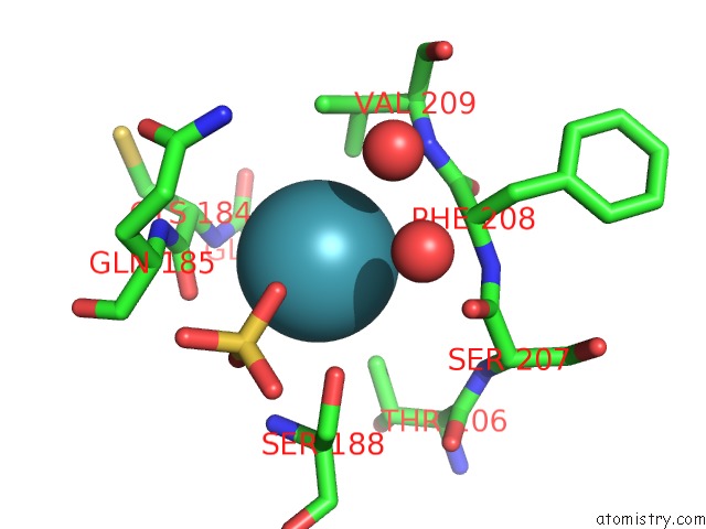
Mono view
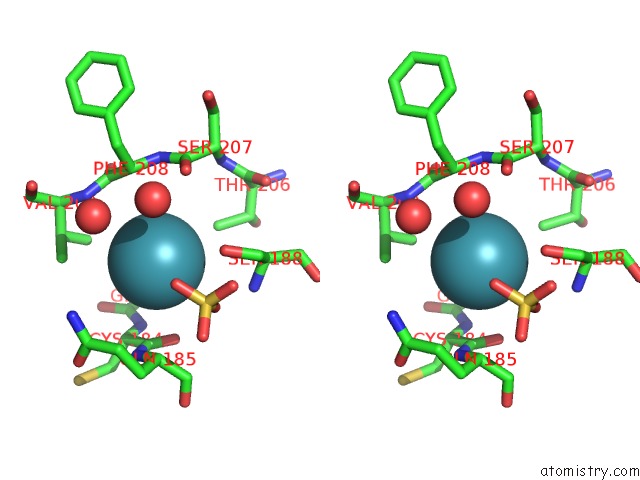
Stereo pair view
Mono view
Stereo pair view
A full contact list of Xenon with other atoms in the Xe binding
site number 1 of On the Routine Use of Soft X-Rays in Macromolecular Crystallography, Part III- the Optimal Data Collection Wavelength within 5.0Å range:
|
Reference:
C.Mueller-Dieckmann,
S.Panjikar,
P.A.Tucker,
M.S.Weiss.
On the Routine Use of Soft X-Rays in Macromolecular Crystallography. Part III. the Optimal Data-Collection Wavelength. Acta Crystallogr.,Sect.D V. 61 1263 2005.
ISSN: ISSN 0907-4449
PubMed: 16131760
DOI: 10.1107/S0907444905021475
Page generated: Sat Oct 12 18:15:25 2024
ISSN: ISSN 0907-4449
PubMed: 16131760
DOI: 10.1107/S0907444905021475
Last articles
Ca in 5S5UCa in 5S5T
Ca in 5S5S
Ca in 5S5R
Ca in 5S5Q
Ca in 5S5P
Ca in 5S5O
Ca in 5S5N
Ca in 5S5M
Ca in 5S5L