Xenon, Xe »
PDB 1c10-1w2z »
1vgi »
Xenon in PDB 1vgi: Crystal Structure of Xenon Bound Rat Heme-Heme Oxygenase-1 Complex
Enzymatic activity of Crystal Structure of Xenon Bound Rat Heme-Heme Oxygenase-1 Complex
All present enzymatic activity of Crystal Structure of Xenon Bound Rat Heme-Heme Oxygenase-1 Complex:
1.14.99.3;
1.14.99.3;
Protein crystallography data
The structure of Crystal Structure of Xenon Bound Rat Heme-Heme Oxygenase-1 Complex, PDB code: 1vgi
was solved by
M.Sugishima,
H.Sakamoto,
M.Noguchi,
K.Fukuyama,
with X-Ray Crystallography technique. A brief refinement statistics is given in the table below:
| Resolution Low / High (Å) | 50.00 / 1.90 |
| Space group | P 32 2 1 |
| Cell size a, b, c (Å), α, β, γ (°) | 65.100, 65.100, 120.500, 90.00, 90.00, 120.00 |
| R / Rfree (%) | 20.1 / 21.7 |
Other elements in 1vgi:
The structure of Crystal Structure of Xenon Bound Rat Heme-Heme Oxygenase-1 Complex also contains other interesting chemical elements:
| Iron | (Fe) | 1 atom |
Xenon Binding Sites:
The binding sites of Xenon atom in the Crystal Structure of Xenon Bound Rat Heme-Heme Oxygenase-1 Complex
(pdb code 1vgi). This binding sites where shown within
5.0 Angstroms radius around Xenon atom.
In total 3 binding sites of Xenon where determined in the Crystal Structure of Xenon Bound Rat Heme-Heme Oxygenase-1 Complex, PDB code: 1vgi:
Jump to Xenon binding site number: 1; 2; 3;
In total 3 binding sites of Xenon where determined in the Crystal Structure of Xenon Bound Rat Heme-Heme Oxygenase-1 Complex, PDB code: 1vgi:
Jump to Xenon binding site number: 1; 2; 3;
Xenon binding site 1 out of 3 in 1vgi
Go back to
Xenon binding site 1 out
of 3 in the Crystal Structure of Xenon Bound Rat Heme-Heme Oxygenase-1 Complex

Mono view
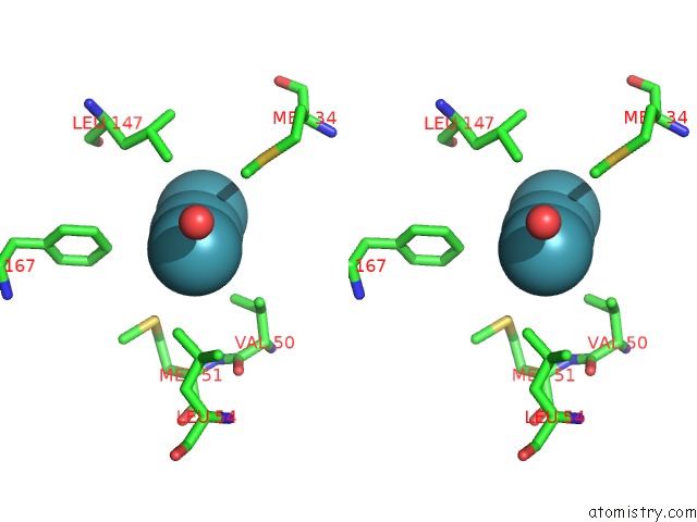
Stereo pair view
Mono view
Stereo pair view
A full contact list of Xenon with other atoms in the Xe binding
site number 1 of Crystal Structure of Xenon Bound Rat Heme-Heme Oxygenase-1 Complex within 5.0Å range:
|
Xenon binding site 2 out of 3 in 1vgi
Go back to
Xenon binding site 2 out
of 3 in the Crystal Structure of Xenon Bound Rat Heme-Heme Oxygenase-1 Complex

Mono view
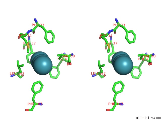
Stereo pair view
Mono view
Stereo pair view
A full contact list of Xenon with other atoms in the Xe binding
site number 2 of Crystal Structure of Xenon Bound Rat Heme-Heme Oxygenase-1 Complex within 5.0Å range:
|
Xenon binding site 3 out of 3 in 1vgi
Go back to
Xenon binding site 3 out
of 3 in the Crystal Structure of Xenon Bound Rat Heme-Heme Oxygenase-1 Complex

Mono view
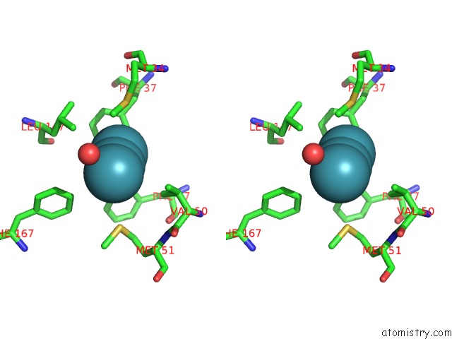
Stereo pair view
Mono view
Stereo pair view
A full contact list of Xenon with other atoms in the Xe binding
site number 3 of Crystal Structure of Xenon Bound Rat Heme-Heme Oxygenase-1 Complex within 5.0Å range:
|
Reference:
M.Sugishima,
H.Sakamoto,
M.Noguchi,
K.Fukuyama.
Co-Trapping Site in Heme Oxygenase Revealed By Photolysis of Its Co-Bound Heme Complex: Mechanism of Escaping From Product Inhibition J.Mol.Biol. V. 341 7 2004.
ISSN: ISSN 0022-2836
PubMed: 15312758
DOI: 10.1016/J.JMB.2004.05.048
Page generated: Sat Oct 12 18:10:00 2024
ISSN: ISSN 0022-2836
PubMed: 15312758
DOI: 10.1016/J.JMB.2004.05.048
Last articles
Zn in 9MJ5Zn in 9HNW
Zn in 9G0L
Zn in 9FNE
Zn in 9DZN
Zn in 9E0I
Zn in 9D32
Zn in 9DAK
Zn in 8ZXC
Zn in 8ZUF